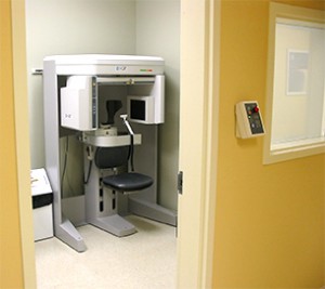
i-CAT Scan
The i-CAT™ is a special type of CT scanner (commonly called a ‘CAT scanner’). Specifically, it is a ‘cone-beam’ CT scanner, unlike most medical scanners , which are ‘spiral CTs’. The advantage of the cone-beam is far less radiation exposure for you than standard medical grade CTs, while maintaining as good, or in some circumstances, even higher resolution. The higher the resolution the more accurate the images.
 The i-CAT™ allows 3-dimensional viewing of your jaws, teeth, and associated structures. From it, anatomy can be visualized and measured very accurately, allowing in many situations better diagnosis than is available on traditional dental x-rays, better planning of potential procedures, and therefore more predictable and efficient surgery, all while minimizing your risk.
You sit in our open-concept machine, positioned in a spacious room where you will be constantly visible to the staff member taking the i-CAT™. Once you are properly positioned, the actual scan takes as little as 5 seconds, and at most less than 30 seconds, during which time you will be asked to remain as still as possible. The scanner is also accessible to patients in wheelchairs. Though considered harmless for use during pregnancy, please notify your surgeon or his staff to discuss the situation.
Depending upon the circumstances, you may be asked to return at a later date to allow your doctor adequate time for manipulation and analysis of the images obtained. This is, of course, for your benefit, and there would be no charge to you for such a follow-up appointment.
In many cases, your dental insurance will provide coverage for part or all of the cost.
The following is a small sample of what can be done with the i-CAT. Not shown are the multiplanar reconstructions, allowing viewing from any angle and 3-D reconstructions of entire faces!
Third Molars (‘wisdom teeth’)
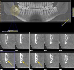 The wide view of all the teeth in the top portion of the image below is similar to a standard panorex radiograph, commonly done at a dental or oral surgery office. It shows an impacted wisdom tooth on the patient’s right side (viewer’s left, shaded in yellow), which does not appear much different than most people of this age. However, in the bottom half of the image, are the cross-sectional slices of the i-CAT in the area of this wisdom tooth (like looking at slices of bread end-on). They show quite clearly that the nerve and blood vessels running inside the jaw bone, called the inferior alveolar neurovascular bundle, are intimately approximated to the root of this tooth, almost wrapped around it! That is illustrated by the small, darker area as indicated by the yellow arrows.
This patient has an elevated risk of nerve injury due to removal of this tooth, possibly resulting in permanent numbness of the lip on this side. This patient may now be informed of this elevated risk, and measures can be taken at the time of surgery to avoid trauma to that specific area.
Dental Implants
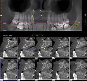 The wide view of all the teeth in the top portion of the image below is similar to a standard panoramic radiograph commonly taken in dental or specialist offices. The area bracketed in yellow is where this patient is missing the four front teeth. There appears on this 2-dimensional view to be sufficient bone height for implants. The question is whether there is enough bone to place a dental implant, or whether adjunctive procedures will be required simultaneously (e.g. small bone graft) or even as a separate procedure months prior to implant placement (e.g. larger bone graft). As demonstrated in the cross-sectional images in the bottom portion of this image, there is clearly NOT enough width of bone, as demonstrated by the narrow bone ridge between the yellow arrows. Superior treatment planning and predictability is possible, reducing the guess-work of having to see and decide at the time of surgery. We don’t like surprises at the time of surgery, and we know you don’t either!
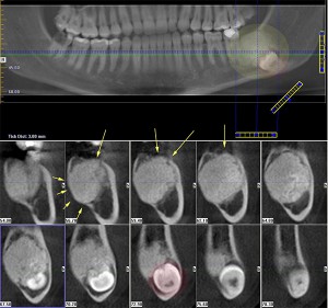
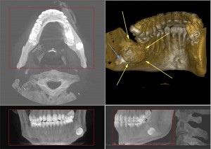
Pathology
The following patient is approximately 20 years old, and the lesion in the lower jaw (highlighted in yellow) was noticed on an x-ray taken to assess his wisdom teeth. The i-CAT allows far superior visualization of the location, size, internal architecture, and effects on the surrounding tissues. In this case, the wisdom tooth has been displaced way down to the very bottom of the jaw (red circle), and the solid mass in his jaw bone can be seen to have expanded beyond the normal confines of the jaw bone (arrows).
This is a 3-D view looking from behind the patient, showing the bulging tumour (yellow arrows) and the tooth at the lower part of the jawbone (blue arrow).
This is a 3-D view looking from behind the patient, showing the bulging tumour (yellow arrows) and the tooth at the lower part of the jawbone (blue arrow).